

Machine shop prints femur model for Fairbanks surgeon
May 31, 2017
Lea Gardine
907-474-7664
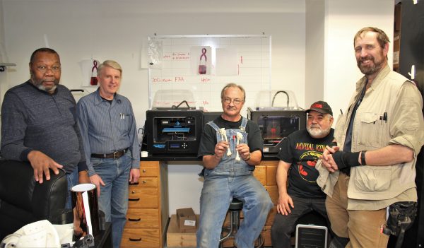
Nothing makes Greg Shipman and his colleagues happier than a challenge.
“We have to be ready for whatever walks through that door,” said Shipman, manager of the machine shop at the University of Alaska Fairbanks Geophysical Institute.
Often, that means creating something mechanical for scientists. Earlier this spring, their work took a more prosthetic turn. A radiologist at Fairbanks Memorial Hospital asked the crew to create a 3-D plastic model of a 12-year-old’s thigh bone, or femur. The model would allow the surgeon assigned to the child’s case to visualize how the surgery would be completed.
For this patient, it was particularly important that the femur be properly aligned. Just a few degrees of rotation could mean the difference between normal knee function and long-term instability of the kneecap. The measurement would have to be very precise — something that is difficult to achieve using two dimensional CT scan images, according to Keir Fowler, the FMH radiologist who worked with Shipman.
“You don’t see the whole bone on any particular image, so it’s hard to do highly precise measurements,” said Fowler. “We also wanted to compare how one femur looked to the other side, so that you can get a nice matching alignment.”
Fowler compared this project to the field of engineering, where scientists frequently have to build prototypes to test their ideas.
“If you have a set of schematics and diagrams that’s all well and good, but to actually determine if a working model is going to function properly, you have to make a model and do demos,” Fowler said.
When Fowler spoke with Shipman, they only had one week before the surgeon was scheduled to operate. To further complicate the process, no one was allowed inside the Elvey Building that weekend due to a construction project. The equipment, the printers and the computers were all moved from the machine shop in the basement of the Elvey Building to the Akasofu Building next door so that the team could get to work creating the model.
The 3-D printers operate in much the same way as CT scans, Shipman said. A bone scan is made up of a series of slices that, when stacked upon each other, make the full bone. The printer builds the bone model by extruding or "printing" layers of thin plastic until the model is complete.
It's a much simpler process than the traditional methods used by machinists, which entails carving a solid chunk of metal or plastic, Shipman said. “With 3-D printing, you still have to design, but you can design almost without limitation — anything you can draw you can print.”
That technology allowed the machine shop team to complete the model in just a few days. The surgeon received it on a Monday — just in time to review the model before the Tuesday morning surgery.
“Think about that — you could help this kid walk properly the rest of his life,” said Jesse Atencio, a technician with the GI’s Research Computing Systems. “The concept is so abstract. You know you’re printing out model of something real that is inside that person’s body … it’s something you can hold and look at all sides of.”
The 3-D printers are mainly used for projects at the Geophysical Institute and UAF in general. However, the possibility of further collaboration with the hospital remains.
“This is the kind of collaboration that makes it really valuable to have resources like this available in a town like Fairbanks,” Atencio said. “Without the university this kind of thing would not be possible.”